

|
|
|
|
|
|
|
|
|
|
|
|
||||||||||||||
|
||||||||||||||
|
|
||||||||||||||
|
||||||||||||||
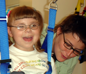 |
Therapy can be fun!
Dr. Stacy Fritz and five-year-old Megan
Howell use a treadmill to improve Megan's gait and motor control. Those
functions were impaired when doctors removed half of Megan's brain to
stop recurring seizures. |
||
|
Posted 08/30/2006 |
|||
|
By Michelle P. Jordan USC Department of Communication Sciences and Disorders 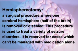 Megan
Howell, 5, is more than ready for kindergarten this fall. She can write
her name, count to 50, and recite her ABCs – pretty impressive for a
child who only has half of her brain. Megan
Howell, 5, is more than ready for kindergarten this fall. She can write
her name, count to 50, and recite her ABCs – pretty impressive for a
child who only has half of her brain.At 15 months of age, Megan underwent a hemispherectomy, a surgery in which the left half of her brain was removed in an effort to stop severe recurring seizures. Aside from a noticeable weakness on the right side of her body that hints at a problem, Megan functions nearly as well as her peers. The question is how? How has the remaining hemisphere of her brain been able to compensate for the loss of skills, such as language, normally controlled by the missing part? How can a better understanding of this process lead to more effective therapy options for future patients?  Researchers at the University of South Carolina’s Arnold School of
Public Health are striving to find out the answers to these questions by
providing federally-funded, cutting-edge rehabilitation services not
offered anywhere else in the nation. Researchers at the University of South Carolina’s Arnold School of
Public Health are striving to find out the answers to these questions by
providing federally-funded, cutting-edge rehabilitation services not
offered anywhere else in the nation.A stroke during birth left Megan suffering from as many as 80 to 100 seizures daily, said her mother, Shelly Howell, of Topeka, Kan. Even though Megan was taking every anti-seizure medication safe for use in children, her condition continued to worsen. By 15 months old, Megan still could neither walk nor talk. Her body and her brain were too occupied with seizures or drugged with medication for there to be room for learning. “She could only sit up. She was like a zombie on the medications,” her mother said. When Megan began to have grand mal seizures that lasted nearly seven minutes, the point at which the brain begins to be deprived of oxygen, Howell and her husband, Jeff, had reached the end of traditional treatment options. Their choice: put Megan on an anti-seizure drug that can be fatal to children, or have half her brain removed. The Howells opted for neurosurgery and in 2002, Megan underwent a hemispherectomy at the University of California Los Angeles. While the seizures stopped immediately, and have not returned since, the difficult road of recovery and rehabilitation was just beginning for the Howells. The two primary hospitals that perform
this surgery, UCLA and Johns Hopkins University, are unable to offer
extended rehabilitation for patients recovering from a hemispherectomy.
USC is beginning to find ways to enhance the long-term rehabilitation of
these patients. De Bode and Dr. Stacy Fritz, a clinical assistant professor in USC’s physical therapy program, are working together to study the brain’s plasticity, its ability to reorganize and reassign neural pathways, after a hemispherectomy. The study is utilizing two promising rehabilitation therapies – Locomotor Training and Constraint-Induced Movement Therapy – to improve participants’ gait and motor control. The goal is to determine which circuits in the brain help perform certain motor tasks.
“We have to understand why,” de Bode said. “No one has ever looked at that before. It’s very technically challenging to study the structural and functional aspects of such a situation because the brain is deformed.” It is due to this degree of difficulty that USC’s state-of-the-art brain imaging equipment, like that found at the McCausland Center, has proven to be invaluable. But it’s not just high-tech equipment and a challenging topic that sets this research and rehabilitation study apart from others. Unlike in many studies, participants here receive a $2,000 stipend to help defray travel and hotel costs, but they also take home something even more precious – functional improvements. It’s a situation in which researchers and
participants both benefit, de Bode said. “If (USC’s research study) helps Megan just the tiniest bit, then it’s worth it,” Howell said. “And if we can help the university better understand hemispherectomies so we can help other families, it’s worth it. Knowledge is power.” For more information: |
|||
 Dr. Stella de Bode, an assistant professor in USC’s Department of
Communication Sciences and Disorders, knew Megan when her brain was
whole and her body was wracked with seizures. De Bode, a former faculty
member at UCLA, is the link that brought Megan and her mother to
Columbia for two weeks this summer to participate in de Bode’s study,
which is funded through a grant from the National Institute of Health.
Dr. Stella de Bode, an assistant professor in USC’s Department of
Communication Sciences and Disorders, knew Megan when her brain was
whole and her body was wracked with seizures. De Bode, a former faculty
member at UCLA, is the link that brought Megan and her mother to
Columbia for two weeks this summer to participate in de Bode’s study,
which is funded through a grant from the National Institute of Health.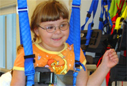
| Columbia, SC 29208 • 803-777-7000 • sphweb@gwm.sc.edu | © University of South Carolina Board of Trustees |